DNA can be extracted from whole blood using a standard phenol/chloroform extraction method. A battery of 25 microsatellite markers which are based on the guidelines of ISAG & FAO’s DAD-IS program can be utilised to generate allelic data. Only forward primers at 5' end of each pair needs labelling with one of the four fluorophores, i.e., FAM (blue), VIC (green), NED (yellow) and PET (red). For details please see Kumar et al., 2009. PCR reactions may be carried out using 50–100 ng genomic DNA in a 25 µl reaction volume.
A touchdown PCR protocol can be used with initial denaturation of 95◦C for 1 min, 3 cycles of 95◦C for 45 s, and 60◦C for 1 min, 3 cycles of 95◦C for 45 s, and 57◦C for 1 min, 3 cycles of 95◦C for 45 s, and 54◦C for 1 min, 3 cycles of 95◦C for 45 s and 51◦C for 1 min, 20 cycles of 95◦C for 45 s, and 48◦C for 1 min with final extension at 72◦C for 5 min. At the end reaction can be stopped by adding 5.0 µl of stop dye (95% formamide, 0.25% bromophenol blue and 0.25% xylene cyanol). 6 µl of PCR products can be loaded on a 2% agarose gel, for electrophoresis to visualize it in UV light after ethidium bromide staining to detect the amplification.
The PCR products can be mixed in a ratio of 1:1.5:2:2 of FAM, VIC, NED and PET labeled primers, respectively. 0.5 µl of this mixture (a post PCR multiplex of 4–5 primers) can be combined with 0.3 µl of Liz 500 as internal lane standard of Applied Biosystems, U.S.A. and 9.20 µl of Hi-Di Formamide per sample. The resulting mixture should be denatured by incubation for 5 min at 95◦C. These denatured samples has to be run on automated DNA sequencer of Applied Biosystems (ABI 3100 Avant). The electropherograms drawn through Gene Scan can be used to extract DNA fragment sizing details using Gene Mapper software (version 3.0) (Applied Biosystems, U.S.A.). |
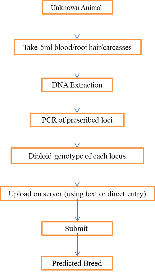
Schematic Diagram
|